
Back to Health Blogs
Health Tips and Blogs

Structure of Kidney - Anatomy, Parts, and Function

5 min read
A
Written ByAdmin
Structure of Kidney - Anatomy, Parts, and Function

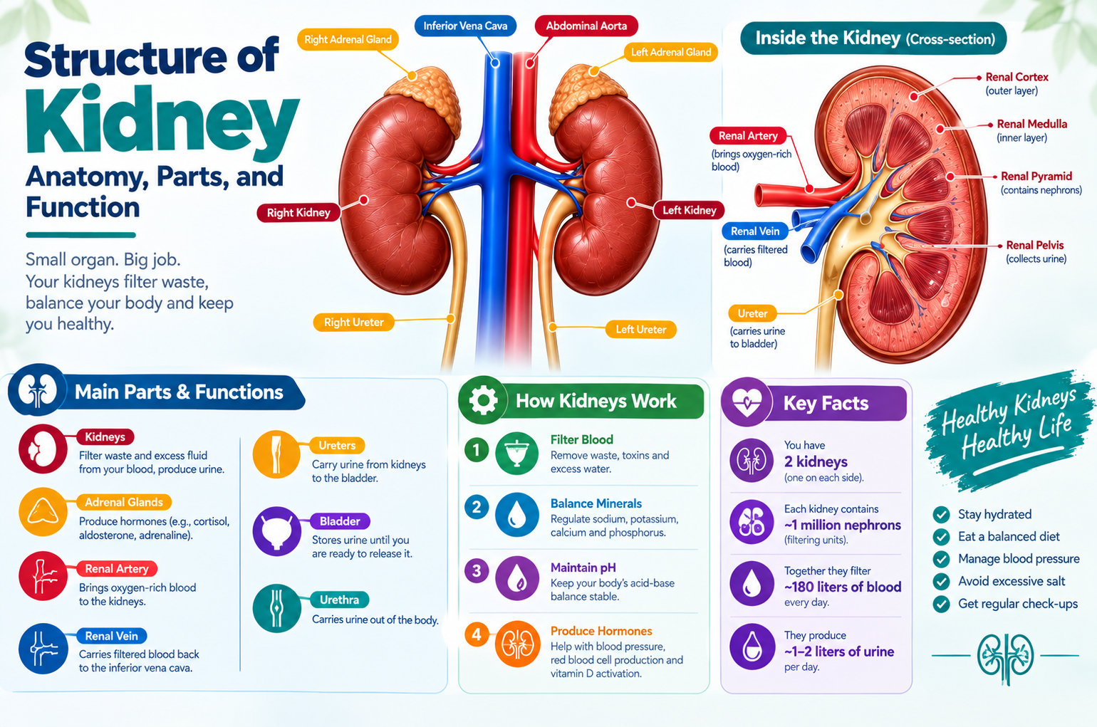
The structure of the kidney reveals one of nature’s most sophisticated filtration systems. Each kidney though small acts as a powerful biological processor, cleansing your blood and maintaining fluid, electrolyte, and pH balance every second of your life.

Understanding kidney structure helps medical students, patients, and healthcare professionals appreciate how this organ’s design directly supports its life-saving functions.

In this guide, we’ll explore the anatomy, parts, layers, and functional units of the kidney, supported by credible medical references like the Mayo Clinic, NIH, and NCBI.

What Is the Kidney?

The kidney is a bean-shaped organ in the urinary system responsible for filtering waste products from blood, producing urine, regulating electrolytes, and controlling blood pressure.

Humans have two kidneys, located on either side of the spine, just below the rib cage.

According to the National Institute of Diabetes and Digestive and Kidney Diseases (NIDDK), each kidney filters around 150 quarts of blood daily, producing about 1–2 quarts of urine.

Location and Size of the Kidney

  • Location: Retroperitoneal (behind the abdominal cavity) on either side of the vertebral column.
  • Right Kidney: Slightly lower than the left due to the liver’s position.
  • Average Size: About 10–12 cm long, 5–7 cm wide, and 3 cm thick.
  • Weight: 120–150 grams in adults (Mayo Clinic).

External Structure of Kidney

Shape, Color, and Weight

  • Shape: Bean-shaped.
  • Color: Dark reddish-brown due to rich blood supply.
  • Surface: Smooth, with a concave medial border and convex lateral border.

Hilum and Capsule Layers

  • Renal Hilum: The entry and exit point for the renal artery, renal vein, lymphatics, and ureter.
  • Capsule Layers:
  1. Fibrous Capsule – Inner layer directly covering kidney tissue.
  2. Adipose Capsule – Fat layer for protection and insulation.
  3. Renal Fascia – Outer connective tissue anchoring kidney to nearby organs.

These layers ensure stability and protect the kidney from physical injury.

Internal Structure of Kidney

When viewed in cross-section, the kidney shows two main regions:

Renal Cortex

  • The outer granular zone of the kidney.
  • Contains renal corpuscles and convoluted tubules.
  • Responsible for initial filtration of blood.

Renal Medulla

  • The inner darker region, arranged into 8–18 renal pyramids.
  • Each pyramid has a base (facing cortex) and apex (renal papilla) pointing toward the renal pelvis.

Renal Pyramids and Columns

  • Renal Pyramids: Cone-shaped structures containing nephron loops and collecting ducts.
  • Renal Columns: Extensions of the cortex between pyramids. They contain blood vessels and connective tissue.

Renal Pelvis, Calyces, and Ureter

  • Minor Calyces: Cup-shaped cavities collecting urine from renal papillae.
  • Major Calyces: Formed by the union of minor calyces.
  • Renal Pelvis: A funnel-shaped structure that collects urine and passes it to the ureter.

Urine produced in nephrons flows from collecting ducts → papilla → calyces → renal pelvis → ureter → bladder.

Nephron – The Functional Unit of the Kidney

Each kidney contains around 1–1.5 million nephrons, which are microscopic structures responsible for filtering blood and producing urine.

Structure of a Nephron

  1. Renal Corpuscle
  • Bowman’s Capsule: A double-walled cup surrounding the glomerulus (capillary network).
  • Glomerulus: Performs ultrafiltration, allowing water and small solutes to pass while retaining proteins and cells.
  1. Renal Tubule
  • Proximal Convoluted Tubule (PCT): Reabsorbs nutrients, water, and electrolytes.
  • Loop of Henle: Maintains a concentration gradient for urine formation.
  • Distal Convoluted Tubule (DCT): Regulates sodium, potassium, and pH balance.
  • Collecting Duct: Drains urine from several nephrons to the renal pelvis.

Types of Nephrons

  • Cortical Nephrons (85%) – Located in the cortex; short loops of Henle.
  • Juxtamedullary Nephrons (15%) – Extend deep into the medulla; crucial for urine concentration.

Blood Supply of the Kidney

The kidneys receive 20–25% of cardiac output—a remarkable amount for such small organs.

Flow Path:

Renal artery → segmental arteries → interlobar arteries → arcuate arteries → interlobular arteries → afferent arteriole → glomerulus → efferent arteriole → peritubular capillaries → renal vein → inferior vena cava

This efficient circulation ensures continuous filtration and nutrient exchange.

Functions Related to Kidney Structure

The kidney’s structure is uniquely adapted to its vital functions:

  • Filtration (via glomerulus)
  • Reabsorption (via tubules)
  • Secretion of waste and toxins
  • Osmoregulation (maintaining water balance)
  • Acid-base balance
  • Blood pressure regulation (via renin-angiotensin system)
  • Erythropoietin production (stimulates red blood cell formation)

Common Disorders Affecting Kidney Structure

  1. Polycystic Kidney Disease (PKD): A genetic condition causing cyst formation.
  2. Glomerulonephritis: Inflammation of the glomeruli affecting filtration.
  3. Hydronephrosis: Swelling due to urine buildup.
  4. Chronic Kidney Disease (CKD): Progressive damage impairing kidney function.
  5. Renal Stones: Solid deposits obstructing urine flow.

The structure of the kidney is a masterpiece of biological engineering—designed for efficiency, precision, and endurance.

From the outer cortex to the inner nephron, each part performs a crucial role in filtering blood, maintaining balance, and supporting life itself.

For medical learners, understanding kidney structure lays the foundation for studying renal physiology and diseases.

If you experience symptoms related to kidney issues (like swelling, fatigue, or urine changes), consult a nephrologist immediately.

Medical Disclaimer

This article is for educational purposes only and is not a substitute for professional medical advice, diagnosis, or treatment. Always consult your doctor or a qualified healthcare provider regarding any medical condition or concerns.

Frequently Asked Questions

Common questions about this health blog answered for you

The main parts include the renal cortex, renal medulla, renal pelvis, and nephrons—each contributing to filtration and urine formation.

The nephron is the smallest structural and functional unit, responsible for producing urine.

Three layers: fibrous capsule, adipose capsule, and renal fascia safeguard the kidney.

Need Medical Consultation?

Our expert doctors are here to help you with personalized care.

Book Appointment
Structure of Kidney - Anatomy, Parts, and Function | Nephro Care India