
Hydronephrosis is a medical condition where one or both kidneys become abnormally swollen due to urine build-up. This occurs when urine flow is obstructed or reverses from the bladder back into the kidneys, creating back pressure that can damage kidney tissue.
In many cases, hydronephrosis develops silently at first. But as urine continues to accumulate, the kidney tissues stretch, often leading to pain, urinary changes, or even infections with symptoms like fever and chills. If left untreated, it can cause permanent kidney damage. Early recognition and timely medical attention are therefore essential to protect kidney function and prevent long-term complications. By understanding this condition, individuals can take proactive steps—seeking care when symptoms arise, monitoring their health, and safeguarding their kidneys.
Understanding the Urinary System
The kidneys, located in the lower back on either side of the spine, filter toxins, waste products, and excess fluids from the blood. These are processed into urine, which travels through narrow tubes called ureters into the bladder—a muscular sac that stores urine until it is released.
In hydronephrosis, this normal urine pathway is disrupted. When the urine fails to drain properly, it backs up in the kidney, causing swelling and discomfort. Although the body can sometimes adapt to minor obstructions, persistent hydronephrosis requires prompt diagnosis and management to avoid lasting damage.
Causes of Hydronephrosis
Hydronephrosis can result from several underlying conditions, including:
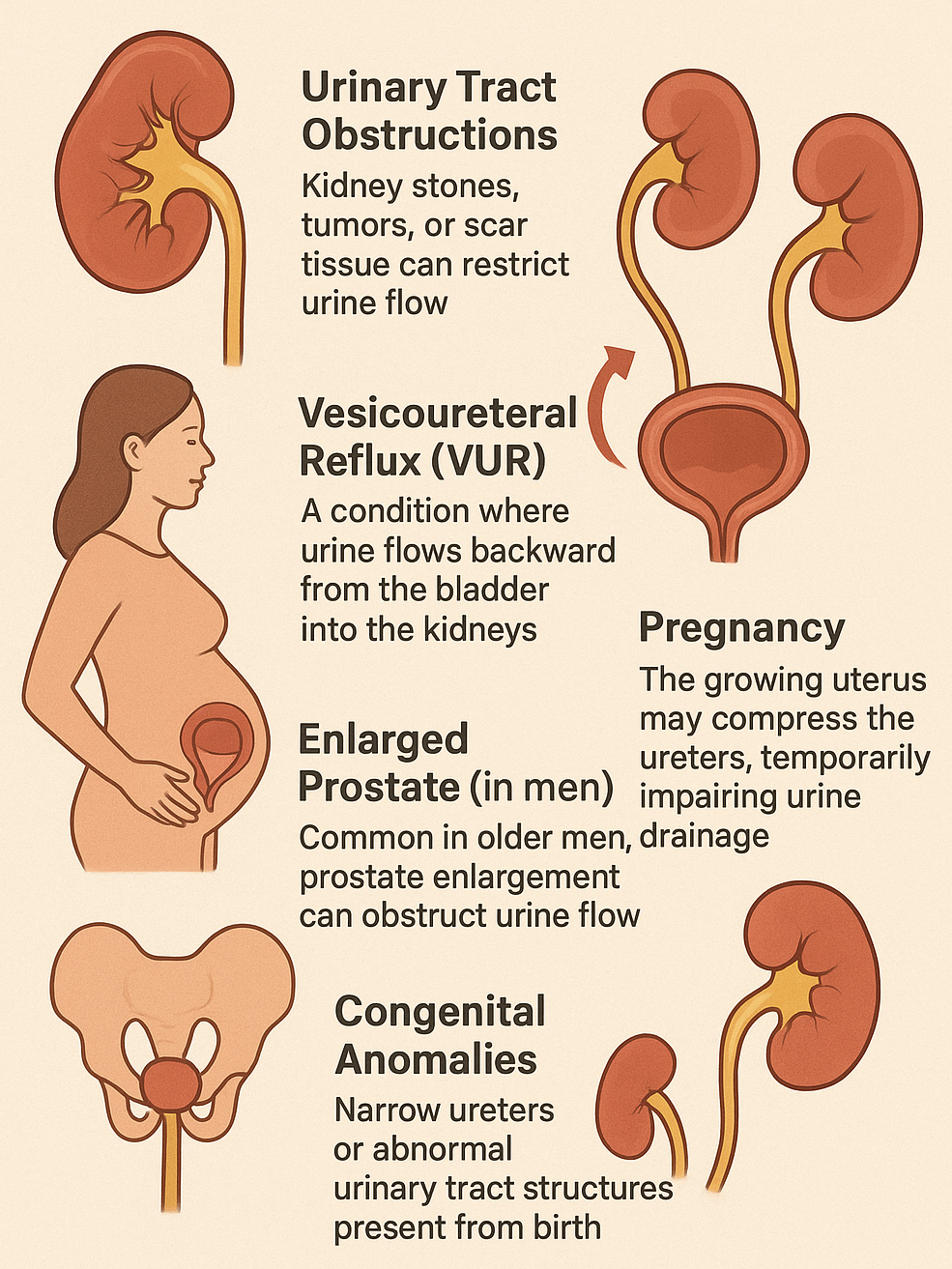
- Urinary Tract Obstructions – Kidney stones, tumors, or scar tissue can restrict urine flow.
- Vesicoureteral Reflux (VUR) – A condition where urine flows backward from the bladder into the kidneys.
- Pregnancy – The growing uterus may compress the ureters, temporarily impairing urine drainage.
- Enlarged Prostate (in men) – Common in older men, prostate enlargement can obstruct urine flow.
- Congenital Anomalies – Narrow ureters or abnormal urinary tract structures present from birth.
Symptoms of Hydronephrosis
The symptoms depend on the severity and speed of progression. Common signs include:
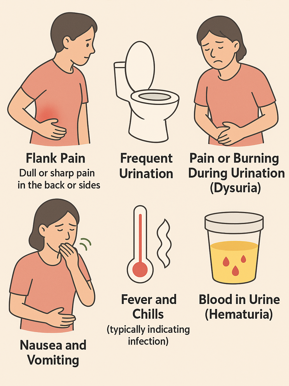
- Flank Pain: Dull or sharp pain in the back or sides.
- Frequent Urination.
- Pain or burning during urination (Dysuria).
- Nausea and vomiting.
- Fever and chills (typically indicating infection).
- Blood in urine (Hematuria).
In some cases, especially mild hydronephrosis, there may be no obvious symptoms, and it is discovered during imaging for other health issues.
Diagnosis
Identifying hydronephrosis involves confirming kidney swelling and finding its underlying cause. The process typically includes:
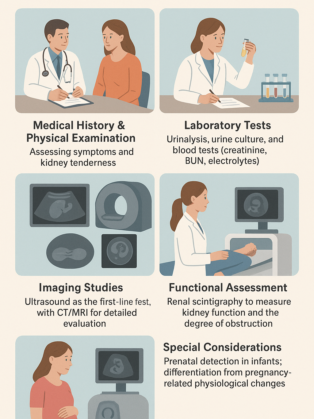
- Medical History & Physical Examination – Assessing symptoms and kidney tenderness.
- Laboratory Tests – Urinalysis, urine culture, and blood tests (creatinine, BUN, electrolytes).
- Imaging Studies – Ultrasound as the first-line test, with CT/MRI for detailed evaluation. In children, VCUG may be used to check for reflux.
- Functional Assessment – Renal scintigraphy to measure kidney function and the degree of obstruction.
- Special Considerations – Prenatal detection in infants; differentiation from pregnancy-related physiological changes.
Treatment Options
The treatment approach depends on the cause and severity:
- Obstruction Relief – Removal of kidney stones, surgery for tumors or structural defects, or prostate treatment.
- Infection Management – Antibiotics for associated urinary infections.
- Drainage Procedures – Placement of a catheter, nephrostomy tube, or ureteral stent to allow urine flow in severe cases.
Possible Complications if Untreated
- Permanent kidney damage
- Chronic kidney disease (CKD)
- Recurrent urinary infections
- Reduced kidney function
Prevention & Self-Care
Not all causes are preventable, but the risk can be reduced by:
- Staying well-hydrated
- Seeking prompt care for urinary symptoms (burning, pain, blood in urine)
- Managing conditions such as kidney stones or prostate enlargement early
Final Thoughts
Hydronephrosis is not a separate disease but a sign of an underlying urinary tract blockage or dysfunction. Diagnosis and treatment should never be delayed, since prolonged obstruction can lead to irreversible kidney injury.
Most patients, however, recover well if the cause is promptly treated—whether through medication, procedures, or surgery. The focus should always remain on protecting kidney function and preventing recurrence.
REFERENCES:
- Babu, R., Venkatachalapathy, E., & Sai, V. (2019). Hydronephrosis severity score: an objective assessment of hydronephrosis severity in children-a preliminary report. Journal of Pediatric Urology, 15(1), 68.e1–68.e6. https://doi.org/10.1016/j. Jpurol.2018.09.020.
- Oliveira, E.A., Oliveira, M.C., & Mak, R.H. (2016). Evaluation and management of hydronephrosis in the neonate. Current Opinion in Pediatrics, 28(2), 195–201. https://doi.org/10.1097/ MOP.0000000000000321.
- Singhal, A., Jain, N., & Chaudhuri, C.R. (2019). A Study of Unilateral Hydronephrosis (A Clinico Radiological-Pathological Correlation) In 100 Consecutive Cases. International Journal of Research, 6, 26-36.
- Zhao, Z. W., Wu, X. C., Deng, J. H., Lian, P. H., & Zhang, X. Bin. (2019). Ureteral obstruction and hydronephrosis caused by foreign body: A case report and literature review. Medicine, 98(44), e17780. https://doi.org/10.1097/MD.0000000000017780.
- Cowan NC. CT urography for hematuria. Nat Rev Urol. 2012;9(4):218–226.
- Doig A, Huether S. Alterations of renal and urinary tract function. In: McCance L, Huether S, eds. Pathophysiology: the biologic basis for disease in adults and children. St. Louis (MO): Elsevier;2014.
- LaRusso, L. (2012, October 1). Hydronephrosis—Adult. Conditions & Procedures, 1-3.
- Unilateral hydronephrosis (2012). US National Library of Medicine. Retrieved from http://www.ncbi.nlm.nih.gov/pubmedhealth/PMH0001535/.